Your heart is a muscle and it pumps blood to your lungs and the rest of your body to supply it with oxygen and nutrients.
Your heart also needs a supply of blood and it gets this from your coronary arteries.
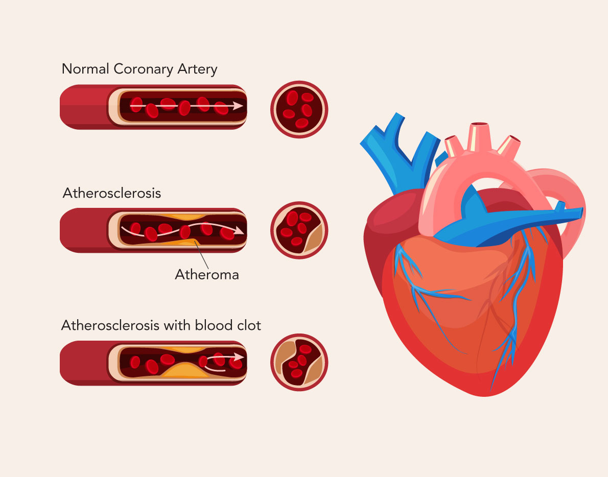
Coronary heart disease happens when cholesterol and other materials build up on the inner surface of your artery walls and form a plaque, which is also called an atheroma. This process of fat building up is called atherosclerosis. It narrows your artery and reduces the blood flow. Thus, the result is your heart muscle does not get all the oxygen it needs. This can damage your heart muscle and lead to a range of symptoms, which can be serious.
Over time, an atheroma may rupture and a clot develops at the rupture site cutting off the blood supply to the heart muscle. This is called a myocardial infarction commonly known as a heart attack.

A heart scan or a coronary calcium scan uses computed tomography (CT) to check for the build-up of plaque on the walls of the arteries of the heart (also known as coronary arteries).
This test checks for the potential narrowing of the coronary arteries in an early stage and determines how severe it is by providing a calcium score. This helps doctors to initiate preventive measures.
- There is no special preparation needed for this test. However, patients need to avoid caffeine and smoking for 4 hours prior to the exam.
- Before the examination, they are required to change into a robe or a gown.
- Metal objects including jewellery, eyeglasses, and hair clips may affect the scan images and should be left at home or removed before the procedure.
- Patients also need to separate their hearing aids and removable dental work.
- Female patients will be asked to remove their undergarments that contain metal underwires.
- Female patients need to inform the technician if they are or could be pregnant.
- The technician will attach sensors called electrodes to the patient’s chest prior to the actual scan. The electrode wires are connected to an electrocardiogram (ECG) and it will record the electrical activity of the patient’s heart.
- The ECG will start recording when the patient’s heart is in the resting stage, which is the best time for the CT (computed tomography) scan to be taken. If the patient’s heart rate is 90 beats per minute or higher, they may be given medication to slow it down.
- During the test, patients will lie on a movable table connected to a CT scanner. The table slides into a round opening of the machine and the scanner will move around the body.
- The CT result does not take long to be produced. Depending on what the images show, the doctor will advise of any lifestyle changes, treatments or procedures that need to be done.
- If the results are normal, they can drive home and continue with their daily activities.


